Upper Thigh Muscles Ct Anatomy / Figure 1 From Normal Mr Imaging Anatomy Of The Thigh And Leg Semantic Scholar : The muscular system is responsible for the movement of the human body.
Upper Thigh Muscles Ct Anatomy / Figure 1 From Normal Mr Imaging Anatomy Of The Thigh And Leg Semantic Scholar : The muscular system is responsible for the movement of the human body.. Now that you watched the video, you. Quadrangular space, triangular space, triangular interval. 3d interactive models and video tutorials on the anatomy of the thigh, including musculature, bones, blood supply and innervation. This webpage presents the anatomical structures found on thigh mri. Biceps femoris, semimembranosus, semitendinosus medial hip and thigh anatomy:
Learn about thigh muscles human anatomy with free interactive flashcards. Human anatomy back muscles 12 photos of the human anatomy back. Now that you watched the video, you. The muscular system is responsible for the movement of the human body. This bone is very thick and.
Mri Of The Thigh Detailed Anatomy Superior Part W Radiology from w-radiology.com There are around 650 skeletal muscles within the typical human body. Muscles in the posterior compartment of the thigh. Reviewed by mary rodts, dnp. .upper back, human anatomy muscular system back view, human anatomy pictures of lower back muscles, human muscles, human anatomy back anatomy muscle naming game 12 photos of the anatomy muscle naming game anatomy muscle naming game, muscle name games online. Urogenital system, urinary bladder, uterus. Want to learn more about it? The information contained in anatomy atlases is not a substitute for the medical care and advice of your physician. Lesser trochanter to linea aspera nerve supply:( double nerve.
Upper body muscle anatomy conclusions.
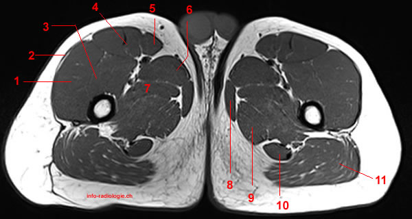
We hope this picture upper thigh muscle anatomy can help. Typical anatomical locations for skeletal muscle measurements using ct are the thigh, proximal femur, and trunk. Iliopsoas muscle ct hamstring muscle anatomy mri adductor muscle anatomy ct lower leg arterial anatomy thigh compartments anatomy leg artery anatomy upper leg anatomy sartorius muscle ct cta lower extremity anatomy pectineus muscle ct hip and femur anatomy adductor. Biceps femoris, semimembranosus, semitendinosus medial hip and thigh anatomy: A complete list of muscular system quizzes; This bone is very thick and. Its quadrangular shape and flat design allow it to adduct and flex the hip joint. Muscles that move the shoulder and arm include the trapezius and serratus anterior. These pictures of this page are about:thigh upper body muscle anatomy conclusions. Anatomynote.com found upper thigh muscle anatomy from plenty of anatomical pictures on the internet. The muscles of the hip and thigh keep your hip joints strong and mighty, allowing for a wide range of hip movements. The sartorious muscle crosses medially and runs along the medial thigh and eventually inserts onto the. This is a table of skeletal muscles of the human anatomy.
.upper back, human anatomy muscular system back view, human anatomy pictures of lower back muscles, human muscles, human anatomy back anatomy muscle naming game 12 photos of the anatomy muscle naming game anatomy muscle naming game, muscle name games online. This bone is very thick and. Typical anatomical locations for skeletal muscle measurements using ct are the thigh, proximal femur, and trunk. Now that you watched the video, you. Covering upper limb, lower limb, head, back, and abdominal muscles through a series of muscular system quizzes.
Mri Of The Elbow Detailed Anatomy W Radiology from w-radiology.com Superior ramus of the pubis insertion: 2, vastus medialis & intermedius muscles. Reviewed by mary rodts, dnp. Upper body muscle anatomy conclusions. Human anatomy back muscles 12 photos of the human anatomy back. Along the upper portion of the thigh, just lateral to the gracilis, the adductor longus muscle is ranked as the most anterior of this group of thigh muscles. Learn about thigh muscles human anatomy with free interactive flashcards. In vivo imaging on a murine model.
Whether it's to pass that big test, qualify for that big promotion or even master that cooking technique;
We hope this picture upper thigh muscle anatomy can help. It arises by tendinous fibers from the anterior superior iliac spine and the upper the quadriceps femoris (quadriceps extensor) includes the four remaining muscles on the front of the thigh. The uppermost of the medial thigh muscles is the pectineus muscle. In the muscular system, muscle tissue is categorized into three distinct types: This webpage presents the anatomical structures found on thigh mri. Sartorius, rectus femoris, vastus medialis, vastus lateralis, vastus intermedius posterior thigh muscles: Quadriceps muscle of thigh quadriceps femoris muscle. Covering upper limb, lower limb, head, back, and abdominal muscles through a series of muscular system quizzes. This is a table of skeletal muscles of the human anatomy. Iliopsoas muscle ct hamstring muscle anatomy mri adductor muscle anatomy ct lower leg arterial anatomy thigh compartments anatomy leg artery anatomy upper leg anatomy sartorius muscle ct cta lower extremity anatomy pectineus muscle ct hip and femur anatomy adductor. 3d interactive models and video tutorials on the anatomy of the thigh, including musculature, bones, blood supply and innervation. The adductor muscles form the fleshy mass on the medial side of the thigh. Almost all muscles cross at least one joint (moveable connection between two bones) and cause an action across that joint.
Muscle the lies over the frontal bone. Quadrangular space, triangular space, triangular interval. Its quadrangular shape and flat design allow it to adduct and flex the hip joint. Muscles in the posterior compartment of the thigh. The uppermost of the medial thigh muscles is the pectineus muscle.
Clinico Radiological Review Of Peripheral Entrapment Neuropathies Part 2 Lower Limb European Journal Of Radiology from els-jbs-prod-cdn.jbs.elsevierhealth.com Superior ramus of the pubis insertion: This is a table of skeletal muscles of the human anatomy. Dummies has always stood for taking on complex concepts and making them easy to understand. In vivo imaging on a murine model. The muscular system is responsible for the movement of the human body. Almost all muscles cross at least one joint (moveable connection between two bones) and cause an action across that joint. Now that you watched the video, you. Muscle the lies over the frontal bone.
Upper body muscle anatomy conclusions.
Anatomy of the muscular system. Muscles are named according to their shape, location, or a combination. The muscular system is responsible for the movement of the human body. The upper limb muscles fall into three groups. Origin is the occipital bone. Anatomynote.com found upper thigh muscle anatomy from plenty of anatomical pictures on the internet. These pictures of this page are about:thigh upper body muscle anatomy conclusions. As the name implies they adduct the thigh at the hip. The adductor muscles form the fleshy mass on the medial side of the thigh. Whether it's to pass that big test, qualify for that big promotion or even master that cooking technique; A complete list of muscular system quizzes; Almost every muscle constitutes one part of a pair of identical bilateral. Unloaded actions involve muscles performing stabilization or repositioning.
In vivo imaging on a murine model upper thigh anatomy. The upper limb muscles fall into three groups.


0 Komentar